1.1 Introduction to Cells
The Cell Theory
Rules of Cell Theory:
-
Living organisms are composed of cells – cells are the building blocks of organisms.
-
Cells are the smallest units of life – a cell is the basic unit capable of carrying out all the functions of a living organism.
-
Cells come from pre-existing cells – cells do not show spontaneous generation (Refer to 1.5 The Origin of Cells).
Additional statements added as microscopes improved:
- Every living cell is surrounded by a membrane
- Cells contain genetic information
- Cells have their own energy release system - powers all of the cell's activities
7 functions all cells carry out for life: Mrs H Gen:
- Metabolism – Living things undertake essential chemical reactions
- Reproduction – Living things produce offspring, either sexually or asexually
- Sensitivity/response – Living things are responsive to internal and external stimuli
- Homeostasis – Living things maintain a stable internal environment
- Growth – Living things can move and change shape or size
- Excretion – Living things exhibit the removal of waste products
- Nutrition – Living things exchange materials and gases with the environment
Exceptions to The Cell Theory
Striated Muscle Cell
Multinucleated Cell: It challenges the idea that a cell has one nucleus, as the muscle cell (fibre) has more than one nucleus per cell (multinucleated). Additionally, the average muscle fibre cell is about 30 mm long, which is much larger than a typical cell.
Giant algae: Acetabularia
Not simple and large: As a single-celled organism, Acetabularia challenges two widely accepted notions about cells: that they must be simple in structure and small in size.
Aseptate fungal hyphae
Multinucleated Cell with a shared Cytoplasm: This challenges the idea that a cell is a single unit as the fungal hyphae have many nuclei, are very large and possess a continuous, shared cytoplasm.
Microscope Magnification
Magnification = size of drawing / actual size
The scale bar represents the actual size of the sample in the image. It can be used to work out the magnification factor or to simply specify the actual length of the specimen or any component within the image.

Cell Differentiation
Specialized tissues can develop by cell differentiation in multicellular organisms
Differentiation involves the expression of some genes and not others in a cell's genome
Stem Cells
Stem cells are unspecialised cells that can give rise to a wide range of body cells by differentiating along different pathways. They retain the capacity to divide indefinitely and have the potential to differentiate into specialised cell types when given the right stimulus.
Types of stem cells:
| Type of Cell |
Differentiated cells produced |
| Totipotent |
Can differentiate into any type of cell including placental cells.
Can give rise to a complete organism. |
| Pluripotent |
Can differentiate into all body cells, but cannot give rise to a whole organism |
| Multipotent |
Can differentiate into a few closely related types of body cell.
|
| Unipotent |
Can only differentiate into their associated cell type. For example, liver stem cells can only make liver cells |
Use of stem cells to treat diseases
Treating Stargardt's disease with stem cells
Stargardt's disease is a disease of the eye. It generally appears in late childhood to early adulthood. It is caused by a recessive genetic mutation in gene ABCA4, which causes an active transport protein on photoreceptor cells to malfunction. This ultimately causes the photoreceptor cells to degenerate.

To treat it, patients are given retinal cells derived from human embryonic stem cells, which are injected into the retina. The results obtained have been quite positive as the inserted cells attach to the retina and become functional, suggesting that it may be possible to restore sight to affected individuals using stem cells.
Treating Leukaemia with stem cells
Leukemia, a type of cancer of the blood or bone marrow, is caused by high levels of abnormal white blood cells. People with leukaemia have a higher risk of developing infections, anaemia and bleeding.
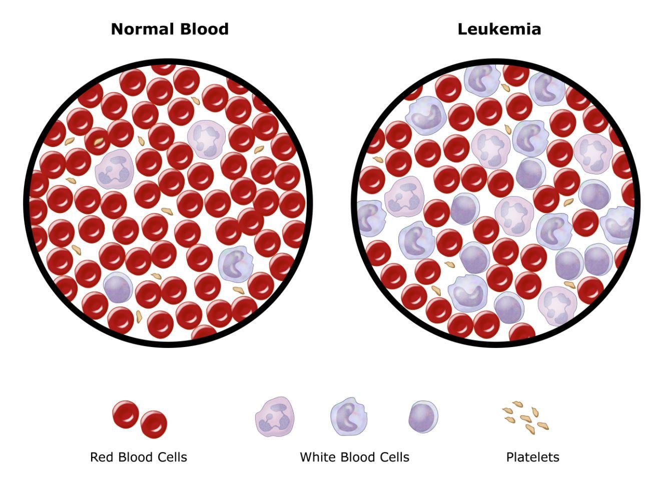
Treatment, in this case, involves harvesting hematopoietic stem cells (HSCs), which are multipotent stem cells. HSCs can be taken from bone marrow, peripheral blood or umbilical cord blood. The HSCs may come from either the patient or from a suitable donor. The patient then undergoes chemotherapy and radiotherapy to get rid of the diseased white blood cells. The next step involves transplanting HSCs back into the bone marrow, where they differentiate to form new healthy white blood cells.
Ethics of Stem Cell Research
| Evaluators |
Embryo |
Umbilical cord blood |
Adult |
| Ethics of extraction |
Involves the death of the embryo which is unethical. |
The umbilical cord is discarded whether or not stem cells are taken from it. |
Does not harm or kill an adult, so it is still ethical |
| Ease of extraction |
Must be attained from an embryo |
Easily attainable and storable |
Difficult to attain as there are very few of them and they are buried deep in tissues |
| Tumour risk |
More risk of becoming tumour cells than others |
Medium chance of malignant tumours developing |
Low chance of malignant tumours developing |
1.2 Ultrastructure of cells
Prokaryotes
They are simple unicellular organisms, with no internal compartmentalisation, no nucleus and no membrane-bound organelles. All metabolic processes thus occur within the cytoplasm. They have 70S Ribosomes.

Cell division in Prokaryotes
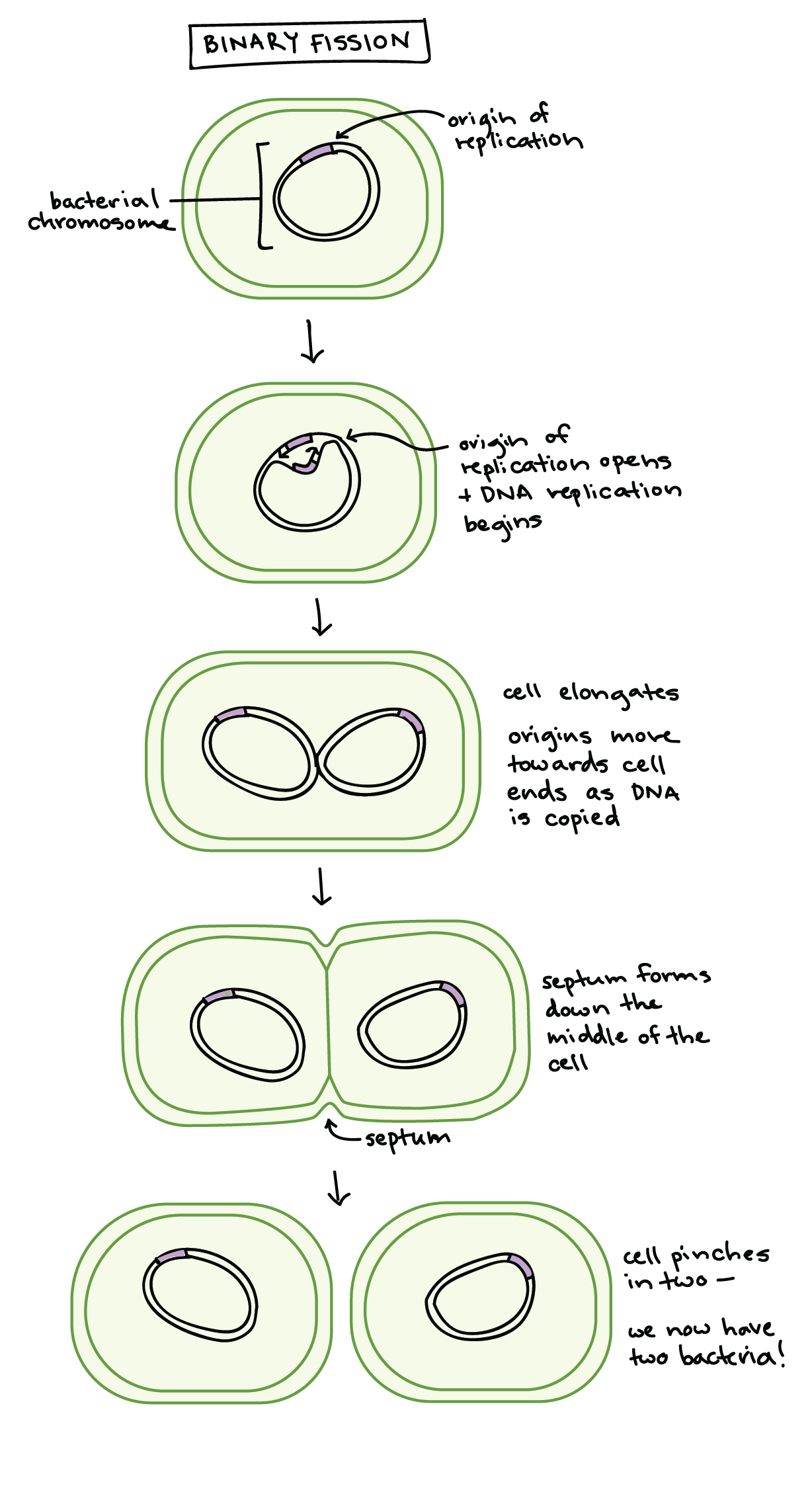
Prokaryotes reproduce by binary fission (a type of cell division) to produce two genetically identical cells.
Eukaryotic cell Ultrastructure
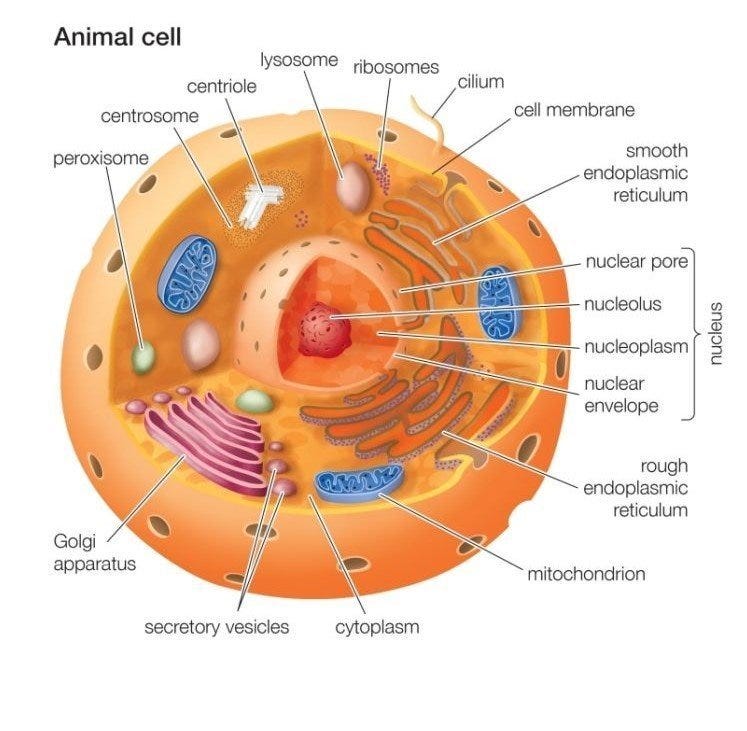
Animal Cell.
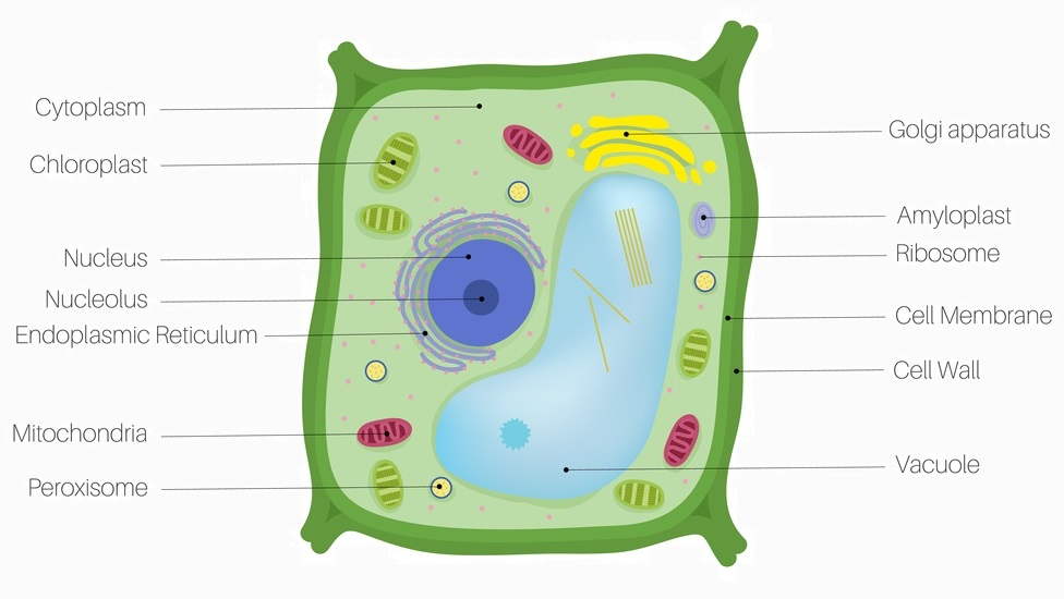
A Plant Cell (palisade mesophyll cell of a leaf)
Electron microscopy
Electron microscopes have a much higher resolution than light microscopes. The resolution of a light microscope is 200 nm compared to 0.1 nm for an electron microscope.
Microscope resolution is the shortest distance between two separate points in a microscope’s field of view that can still be distinguished as distinct objects.
It can differentiate objects as small as 0.001 µm. It played a part in understanding the structure of organelles, such as the thylakoid membranes of chloroplasts, and the actin and myosin filaments of muscle.
Eukaryotic cells can be observed using a light microscope. At this magnification, only larger structures such as cell membrane, cell wall, nucleus, central vacuole and chloroplasts can be seen.
For a detailed view of cells, an electron microscope is usually used.
1.3 Membrane structure
Phospholipid bilayers

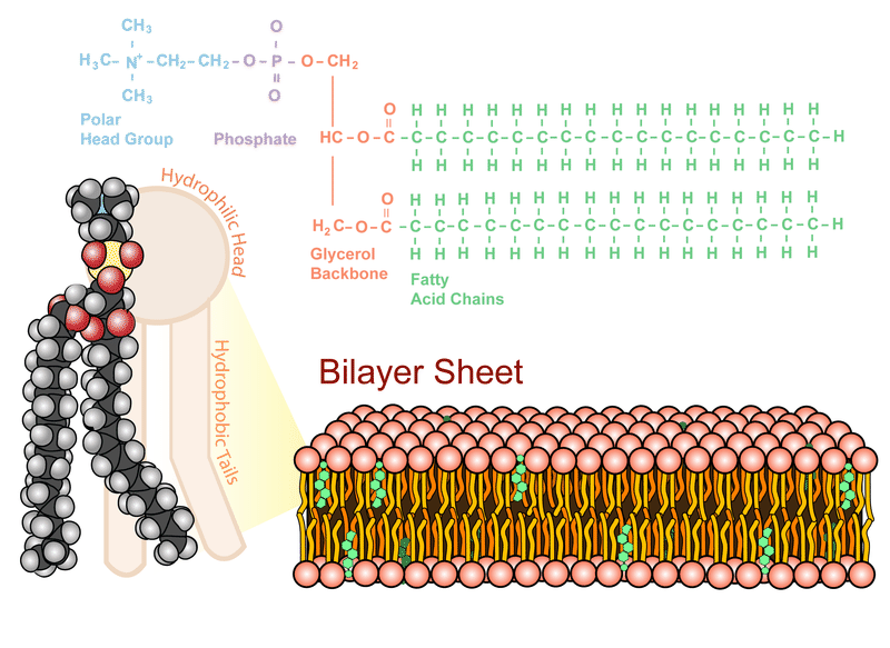
The Fluid Mosaic Model

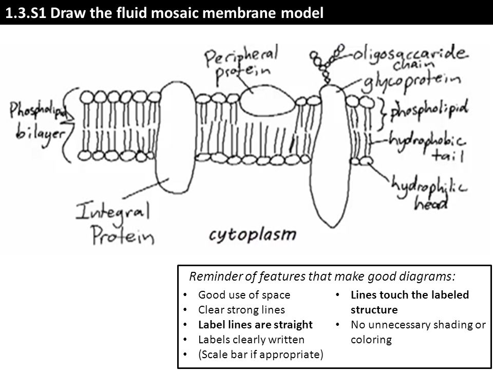
Typical exam drawing question:
Cholesterol in membranes
Cholesterol is a type of liquid, but is not a fat or oil. It belongs to the group called steroids.
Its role in the phospholipid bilayer:
- Cholesterol forms an integral part of an animal membrane and plays an important role in controlling membrane fluidity and permeability to some solutes.
- The cholesterol in the membrane restricts the movement of phospholipids and other molecules, thus reducing membrane fluidity.
- At low temperatures, it disrupts the regular packing of the hydrocarbon tails of phospholipid molecules, which prevents the solidification of the membrane. This enables the membrane to stay more fluid at lower temperatures, allowing the membrane to function properly.
- It reduces membrane permeability to hydrophilic molecules and ions such as sodium and hydrogen.
1.4 Membrane transport
Endocytosis:
The fluidity of membranes allows materials to be taken into cells by endocytosis
Passive methods of transport across membranes
Simple diffusion
Simple diffusion occurs in a gas or liquid medium and only requires a concentration gradient. Simple diffusion across membranes involves particles passing between the phospholipids in the membrane. It can only happen if the phospholipid bilayer is permeable to the particles. Non-polar particles such as oxygen can diffuse through easily.
Facilitated diffusion
Ions and particles that cannot diffuse between phospholipids can pass into or out of cells if there are channels for them through the plasma membrane. Because these channels help particles to pass through the membrane, from a higher concentration to a lower concentration, the process is called facilitated diffusion.

Osmosis
Water is able to move in and out of most cells freely. This net movement is osmosis.
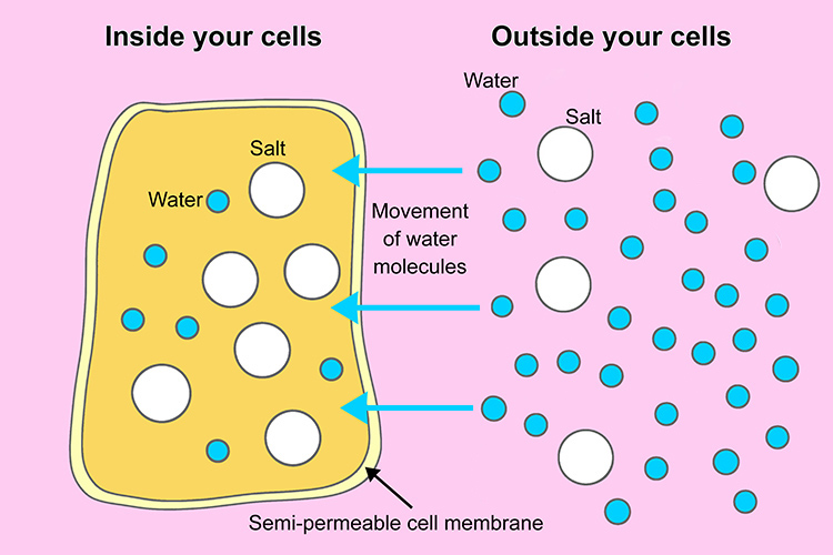
Osmolarity refers to the concentration of a solution in terms of moles of solutes per litre of solution.
1.5 The Origin of Cells
Pasteur's Experiment
Louis Pasteur (1822–1895), a famous French microbiologist, gave crucial evidence to support the hypothesis that cells must come from pre-existing cells. His experiment disproved the theory of spontaneous generation, which stated that life could appear from a combination of dust, air and other factors.

Miller–Urey experiment
Miller and Urey recreated the conditions of early Earth in a closed system by including a reducing atmosphere (low oxygen) with high radiation levels, high temperatures and electrical storms. After running the experiment for a week, some simple amino acids and complex oily hydrocarbons were found in the reaction mixture. This experiment proved that the non-living synthesis of simple organic molecules was possible.

The Endosymbiotic Theory
The theory of endosymbiosis helps to explain the evolution of eukaryotic cells. It states that mitochondria were once free-living prokaryotic organisms that had developed the process of aerobic cellular respiration. Larger prokaryotes that could only respire anaerobically took them in by endocytosis. They allowed them to live in their cytoplasm. As long as the smaller prokaryotes grew and divided as fast as the larger ones, they could persist indefinitely inside the larger cells. The cells evolved to develop a mutualistic relationship turning it into a eukaryote with membrane-bound organelles. In plant cells, the same thing happened with chloroplasts. If a prokaryote that had developed photosynthesis was taken in by a larger cell and was allowed to survive, grow and divide, it could have developed into the chloroplasts of photosynthetic eukaryotes. 
1.6 Cell division
Cell cycle

Cyclins

Mitosis

Cytokinesis
It is the division of the parental cytoplasm between the two daughter cells after mitosis (though it often starts in telophase).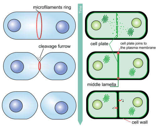
Chromatography Review
Chromatography: A technique for separating the components, or solutes, of a mixture on the basis of the relative amounts of each solute distributed between a moving fluid stream, called the mobile phase, and a contagious stationary base. The mobile phase may be either a liquid or a gas, while the stationary phase is either a solid or a liquid

View count: 17739